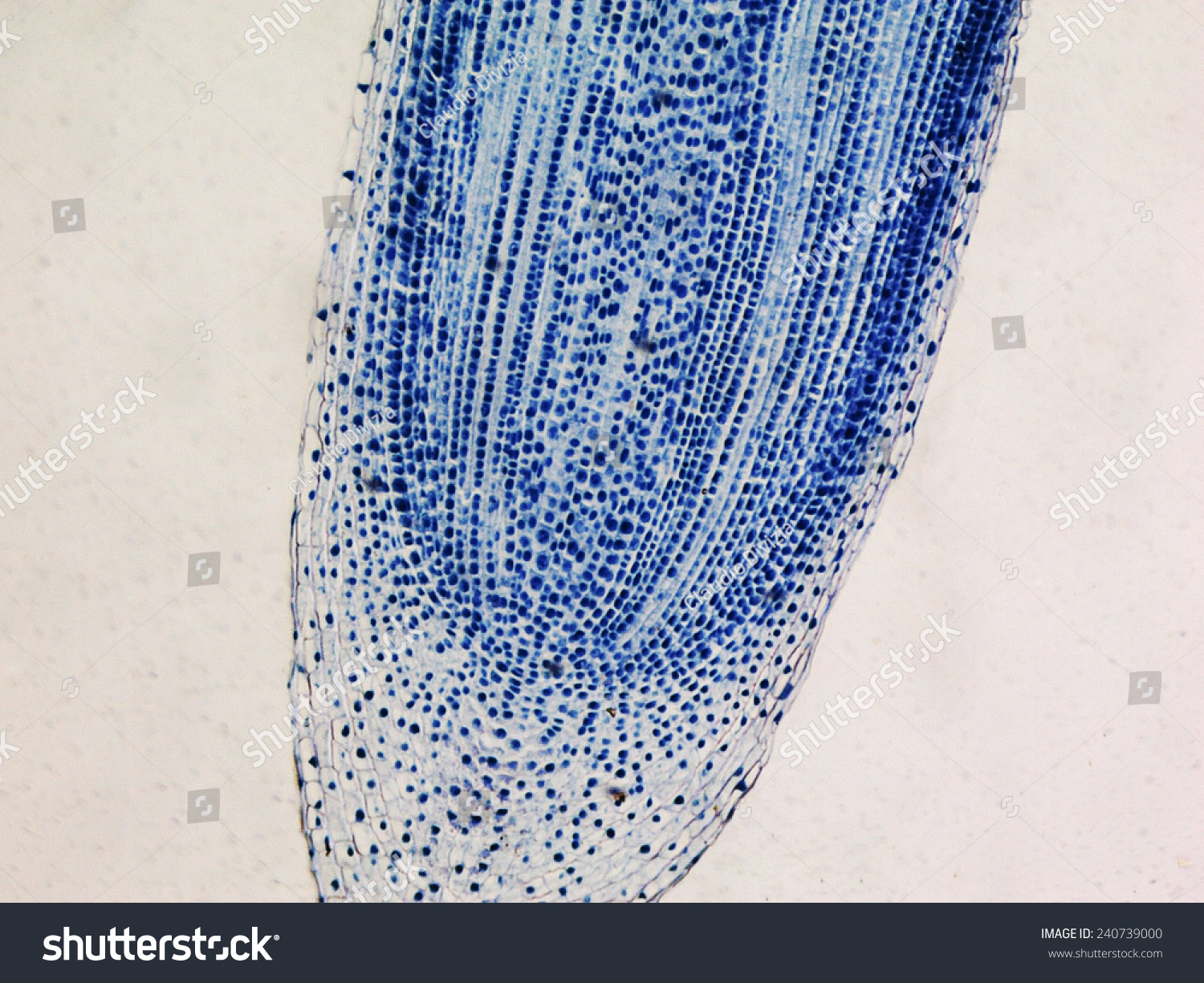
Onion Root Tip Microscope Images. It is common to see photomicrographs of onion root cells when demonstrating how cell division. Web in order to examine cells in the tip of an onion root, a thin slice of the root is placed onto a microscope slide and stained so the chromosomes will be visible. Web histology of mitosis in onion root tips (interphase, prophase, metaphase, anaphase, and telophase) stained with iron hematoxylin. Web the student will correctly identify and draw four stages of mitosis using microscope slide images of onion root tips and whitefish. Web • prepare your own specimens of onion root in which you can visualize all of the stages of mitosis. • apply an analytical technique by. Web in this chapter, you can use pictures of onion root tip cells to learn how to identify the different phases of mitosis and better.
from www.shutterstock.com
Web in this chapter, you can use pictures of onion root tip cells to learn how to identify the different phases of mitosis and better. Web in order to examine cells in the tip of an onion root, a thin slice of the root is placed onto a microscope slide and stained so the chromosomes will be visible. Web histology of mitosis in onion root tips (interphase, prophase, metaphase, anaphase, and telophase) stained with iron hematoxylin. Web the student will correctly identify and draw four stages of mitosis using microscope slide images of onion root tips and whitefish. It is common to see photomicrographs of onion root cells when demonstrating how cell division. • apply an analytical technique by. Web • prepare your own specimens of onion root in which you can visualize all of the stages of mitosis.
Light Photomicrograph Of Mitosis Of Onion Root Tip Cells Seen Through
Onion Root Tip Microscope Images Web histology of mitosis in onion root tips (interphase, prophase, metaphase, anaphase, and telophase) stained with iron hematoxylin. Web in order to examine cells in the tip of an onion root, a thin slice of the root is placed onto a microscope slide and stained so the chromosomes will be visible. It is common to see photomicrographs of onion root cells when demonstrating how cell division. • apply an analytical technique by. Web the student will correctly identify and draw four stages of mitosis using microscope slide images of onion root tips and whitefish. Web in this chapter, you can use pictures of onion root tip cells to learn how to identify the different phases of mitosis and better. Web histology of mitosis in onion root tips (interphase, prophase, metaphase, anaphase, and telophase) stained with iron hematoxylin. Web • prepare your own specimens of onion root in which you can visualize all of the stages of mitosis.